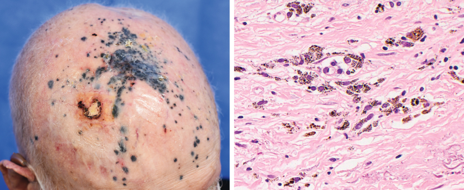
An 84-year-old man with a history of melanoma of the right forehead complicated by in-transit metastases to the scalp presented to the dermatology clinic. Seven months prior, treatment with two immunotherapy agents had been started, which had resulted in shrinking of the metastatic scalp nodules. On physical examination, there were multiple blue-gray macules on the scalp, as well as an unhealed ulcer from trauma. Dermoscopy-guided biopsy of four macules revealed scattered melanin-laden macrophages within the superficial and middle dermis without any evidence of residual melanoma. Immunohistochemical staining of a biopsy sample for markers of melanoma was also negative. What is the most likely diagnosis?一位84岁男性,右额部黑色素瘤病史,伴有头皮的途中转移,前来皮肤科就诊。7个月前,他开始接受两种免疫治疗药物的治疗,这使得转移性头皮结节有所缩小。体格检查发现头皮上有多个蓝灰色斑点,以及一个因外伤导致的未愈合溃疡。在皮肤镜引导下对四个斑点进行活检,结果显示在表皮和中层真皮内有散在的含黑色素的巨噬细胞,但未发现任何残留黑色素瘤的证据。活检样本的免疫组化染色也未检测到黑色素瘤标志物。最可能的诊断是什么?
A. Lentigo maligna恶性黑素瘤
B. Metastatic melanoma转移性黑色素瘤
C. Post inflammatory hyperpigmentation炎症后色素沉着
D. Solar lentigo日光性雀斑
E. Tumoral melanosis肿瘤性黑变病
答案解析:
肿瘤性黑变病(Tumoral melanosis)是一种色素性皮肤病变,当黑色素瘤细胞释放的黑色素被真皮层的巨噬细胞吞噬时就会出现这种情况。它通常在黑色素瘤消退后出现,有时是免疫治疗的反应。病变部位缺乏结节性表现支持肿瘤性黑变病的诊断,但需要通过组织学评估来区分黑色素瘤转移和肿瘤性黑变病。
在组织学上,肿瘤性黑变病表现为真皮或皮下组织中大量聚集的含黑色素的巨噬细胞(黑色素巨噬细胞),这些细胞呈结节状或片状排列。这种病变通常缺乏活的黑色素细胞,免疫组化染色也通常为阴性。因此,在临床和组织学检查中,需要仔细区分肿瘤性黑变病与黑色素瘤转移。
正确答案:E
原创文章(本站视频密码:66668888),作者:xujunzju,如若转载,请注明出处:https://zyicu.cn/?p=19809
 微信扫一扫
微信扫一扫  支付宝扫一扫