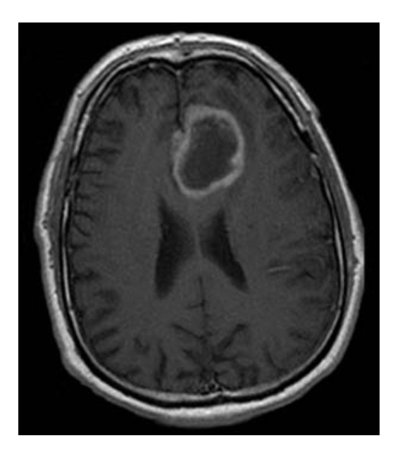
A 41-year-old man visits his physician because of increasingly painful headaches. CT of the head reveals an abnormality and an MRI of the brain with contrast (shown below) is performed for further evaluation. If a biopsy of this tumor were obtained, what would the pathologist likely see under the microscope?一名 41 岁的男子因头痛加剧而去看医生。 头部 CT 显示异常,并进行脑部 MRI 对比(如下所示)以进行进一步评估。 如果对该肿瘤进行活检,病理学家可能会在显微镜下看到什么?
(A) Densely packed cells with halos of cytoplasm surrounding large round nuclei
(B) Perivascular pseudorosettes with tumor cells surrounding vessels
(C) Pseudopalisading tumor cells surrounding necrotic regions
(D) Sharply demarcated areas of tumor cells located at the grey-white matter junction
(E) Whorled pattern of concentrically arranged spindle cells with psammoma bodies
答案解析:
该患者的病变与多形性胶质母细胞瘤 (GBM) 一致,GBM 是最常见的原发性脑肿瘤。 MRI 显示一个不规则的肿块病变,周围有强烈的对比增强和中央低增强,这是由于中央坏死提示高度恶性肿瘤。 请注意病变的“蝴蝶”形状,因为它在穿过中线时通过胼胝体时变窄——这是 GBM 中的经典影像学发现。 在组织病理学上,病变由围绕坏死区域的高度恶性的星形胶质细胞肿瘤细胞组成; 这被称为伪栅栏。 GBM 与遗传改变有关,包括 p53 功能丧失、表皮生长因子受体基因 (EGFR) 活性增加以及染色体臂 10q 杂合性丧失。 GBM 预后差,诊断后平均生存期为 8-10 个月; 大多数患者在两年内死亡。
正确答案:C
原创文章(本站视频密码:66668888),作者:xujunzju,如若转载,请注明出处:https://zyicu.cn/?p=14721
 微信扫一扫
微信扫一扫  支付宝扫一扫